|
If you have Tibialis Posterior Tendonitis, be aware that there is nothing deficient about your
Tibialis posterior tendon or muscle....
The real problem is that the
Posterior Tibial tendon and muscle are being overloaded by
faulty patterns of
leg alignment and muscle tensioning.
If Tibialis Posterior Rehab is to work, you must correct these things.
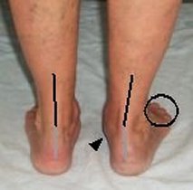
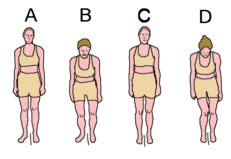
(<<< See Alignment Faults for Explanation of Picture)
|

About the Author...

Why donate?
PayPal Membership No Longer Required!
|
|

Table of Contents
Page 1: Tibialis Posterior Tendonitis Rehab: Introduction
(<<< You are here)
Page 2: Posterior Tibial Insufficiency Treatment (1): Leg Realignment
Page 3: Posterior Tibial Insufficiency Treatment (2): Exercises
Page 4: Posterior Tibial Tendonitis Treatment: Trigger Point Massage
Page 5: Posterior Tibial Insufficiency Treatment
(4): Postural Realignment Made Easy!
On this Page:
Posterior Tibialis Pain Symptoms
Posterior Tibialis Anatomy
Posterior Tibialis Action
Posterior Tibialis Alignment Faults
Posterior Tibialis Pain Symptoms
Trigger points in the Posterior Tibial muscle cause pain down the back of the calf (1),
which may extend down over the Achilles tendon and the back of the heel. Often pain is
worst upon standing first thing in the morning.
Tibialis Posterior tendon pain (and possibly swelling) is to be felt along the tendon
as it curves around the back of the knob on the inside ankle (medial maleolus) (2).
There may also be an ache that extends into the foot arch(2). Again, symptoms
are likely to be worst first thing out of bed.
Posterior Tibialis Anatomy
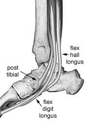
| The posterior tibial muscle starts from the back of the tibia (hence its name!), its
tendon descends behind and then under the medial maleolus (the bump on the inside of
the ankle), and then inserts into the bones of the under-foot just in front of the ankle.
The muscle fibers are arranged in pennate fashion (like a bird's feather), which means
that it is very strong, but has only small range of functional movement. That small range
of functional movement is a significant factor in the pathology of the Tibilais posterior.
As soon as you attempt to work it outside
of its functional range, it works at a fraction of its mid range strength, and is easily damaged. |
Diagram 1 : The Tibialis posterior looking from the
back (posterior view). Note the "pennate" arrangement of the muscle fibers.

|
Diagram 2: The Tibialis posterior viewed from inside of the ankle (medial view).
|
Posterior Tibialis Action
The Tibialis posterior raises the instep and rotates the foot medially such that the
toes are pointing inwards ("pigeon toed stance"). In anatomical terms, this is inversion
and medial rotation of the foot. Medical texts state that the foot is plantar flexed as
well, but this action is a weak one, and it will be ignored in the discussion that follows.
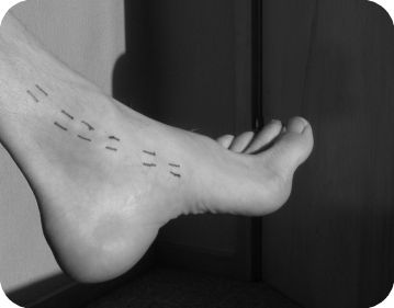
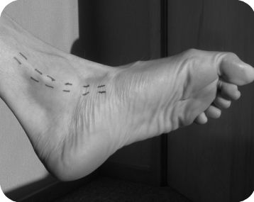
Pictures: The action of the Tibialis posterior. The lines marked in ink delineate the
Tibialis posterior tendon.
Picture 3: The Tibialis posterior is lengthened. The instep is lowered, and the foot is
laterally rotated (toes pointing away from the center line).
|
Picture 4: The Tibialis posterior is contracted. The instep is raised, and the toes
are medially rotated.
Note also the extension and flexion of the toes. Toe movement is not a function of
the posterior tibial muscle, but flexing the toes does aid Tibialis posterior in its arch lifting action.
|
Similar descriptions could be found in any medical text book, but are only a starting point
in our search for the real cause and the real solution. We need to go further.
Posterior Tibialis Alignment Faults
Alignment faults around the ankle are associated with an over-elongated Tibialis posterior.
The inside of the ankle drops, the foot arch flattens, a line drawn through the Achilles
tendon and back of heel bows inward. The toes point outwards such that when the kneecap is pointing forward the
toes point to the side: The foot is rotated laterally with reference to the knee.
Alignment faults around the knee are associated with a knock kneed stance
(the femur rotates medially and adducts causing the knee cap of
the affected leg to "look inward"). See diagram 6 for further discussion.
|
|
Diagram 5: Posterior Tibialis Tendonitis pictured from the rear. The inside of the
ankle is dropped (arrow), the foot
arch is flattened, and a line drawn through the Achilles tendon and back of heel has bowed inward.

|
Diagram 6: Posterior Tibialis Tendonitis & Inward collapsing Knee.
A and B: When the patient is asked to point the knee cap straight ahead, the knee
travels directly forward during the squatting movement, but all the weight is over
the inside (medial) edge of the foot, which causes the foot arch to collapse.
|
|
|
| By
itself, the Tibialis anterior muscle is not strong enough to prevent this happening. C and D: When the patient is asked to point the foot straight ahead to correct the
faulty alignment between the knee and the foot, this alignment does not change.
The only thing that changes is that the thigh bone rotates inward and adducts into
the "knock kneed stance" displayed in "D".
|
|
|
|
Return to top...

Tibialis-posterior rehabilitation © Bruce Thomson EasyVigour Project
|